Lab notebook
Investigating Collagen-Induced Fibrin Fibrillogenesis in the Absence of Thrombin
Day 0 Fri 7/10/26
- Read literature & discuss
- Coradin, Wang, Law & Trichet (2026): Review of collagen-fibrin mixed hydrogels
- Cummings, Gawlitta, Nerem & Stegemann (2004): Engineered vascular constructs
- Hansen et al. (2026): Unraveling the molecular mechanism of in situ surface-initiated thrombogenesis
Day 1 Mon 7/13/26
- Read literature & discuss
- Luo et al. (2020): Dental pulp stem cell mineralization on collagen-fibrin scaffolds
- Lai, Frey, Kerandi, Lake, Tranquillo & Barocas (2012): Microstructural/mechanical differences in co-gels
- Kim, Litvinov, Chen, Chen, Weisel & Alber (2017): Compression behavior of fibrin-collagen composites
Day 2 Tue 7/14/26
- Sliced silicon [100] wafers into ~1 cm × 1 cm pieces
- Collagen solution prep (Shifu)
- Solute: type I telocollagen, only soluble in acid; stock received at pH 4
- Neutralized to pH 7 with NaOH so collagen comes out of solution onto the wafer
- Diluted to 5 µg/mL and 50 µg/mL
Day 3 Wed 7/15/26
- Deposited collagen thin films: wafers left in 5 and 50 µg/mL collagen for 18 h
Day 4 Thu 7/16/26
- Rinsed collagen wafers (collagen-only procedure)
- Aspirate collagen/PBS solution
- Cover wafers with PBS and aspirate, ×3
- Fibrinogen dilution calc: 100 µg/mL → mix 290 mL PBS with 7.5 mL fibrinogen stock (FS)
- Deposited 4 mg/mL fibrinogen on 5 µg/mL collagen wafers, 18 h adsorption
Day 5 Fri 7/17/26
- Rinsed fibrinogen-on-collagen wafers
- PBS ×3, then DI water ×3
- Aspirated all water off to prep for AFM
- AFM: 4 mg/mL fibrinogen, 18 h, on 5 µg/mL collagen
- Large, irregular fibres (~500 µm) formed on collagen without thrombin
- Optical follow-up of the same sample on 7/18
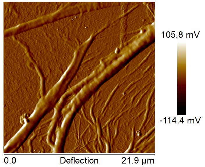
AFM, 21.9 µm scan: thick fibres several µm wide cross the collagen film, with finer fibrils branching off them. 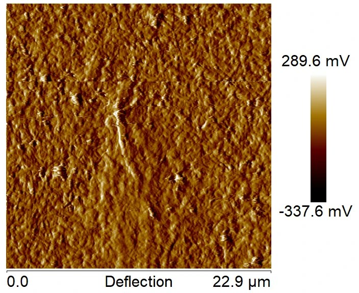
AFM, 22.9 µm scan of the same 4 mg/mL, 18 h sample: away from the large fibres, the surface is a rough, densely textured protein layer. 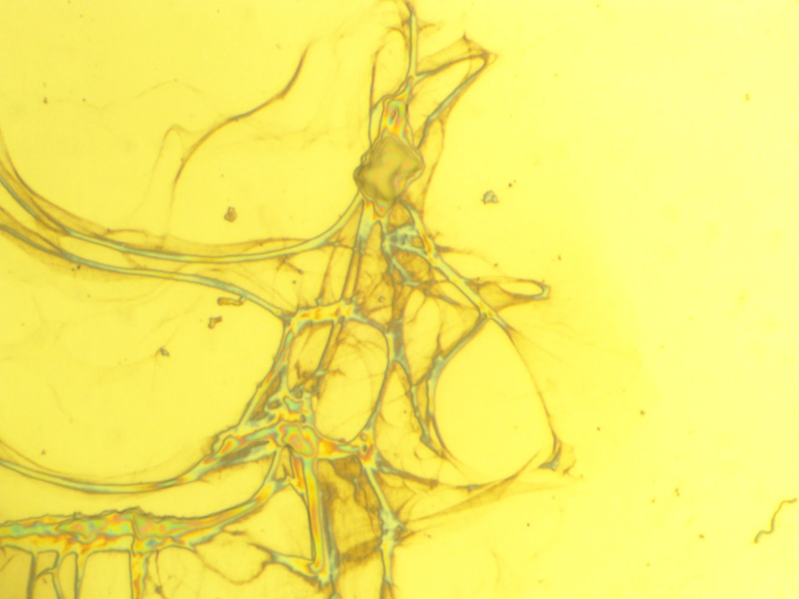
Optical, 10×: 4 mg/mL fibrinogen after 18 h on 5 µg/mL collagen. A branching network of large fibres formed on the collagen film with no thrombin added. - Deposited 100 µg/mL fibrinogen on 5 µg/mL collagen for 18 h adsorption
Day 6 Mon 7/20/26
- AFM: collagen-only control, 5 µg/mL (negative control for fibrin–collagen images)
- AFM: 100 µg/mL fibrinogen, 18 h, on 5 µg/mL collagen
- No fibres comparable to the 4 mg/mL sample → fibre formation looks concentration-dependent
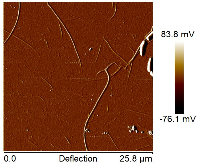
AFM, 25.8 µm scan: at 100 µg/mL there are only thin, scattered fibrils, nothing like the thick fibres at 4 mg/mL. - Deposited 100 µg/mL and 4 mg/mL fibrinogen on 50 µg/mL collagen, 18 h
Day 7 Tue 7/21/26
- Optical: fibrinogen, 18 h, on 50 µg/mL collagen
- 4 mg/mL vs 100 µg/mL fibrinogen
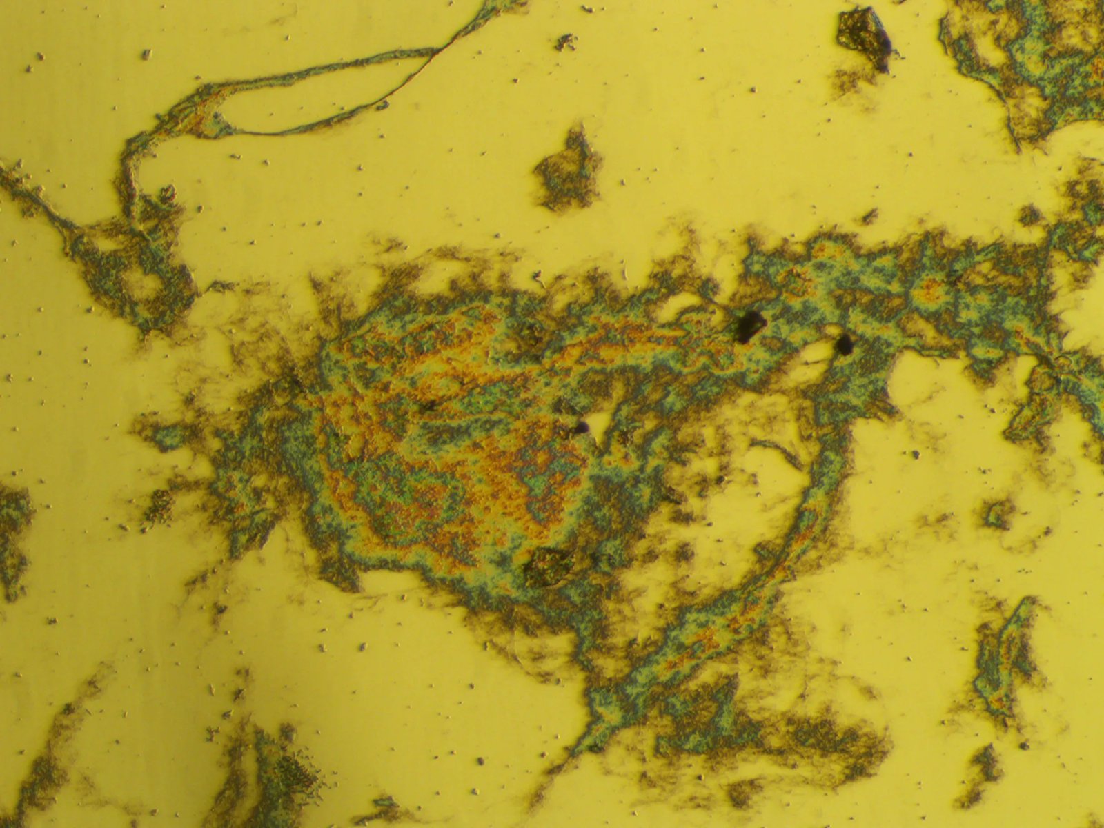
Optical, 10×: 4 mg/mL fibrinogen, 18 h on 50 µg/mL collagen. Thick, irregular aggregates (rainbow thin-film colours) joined by fibrous strands. 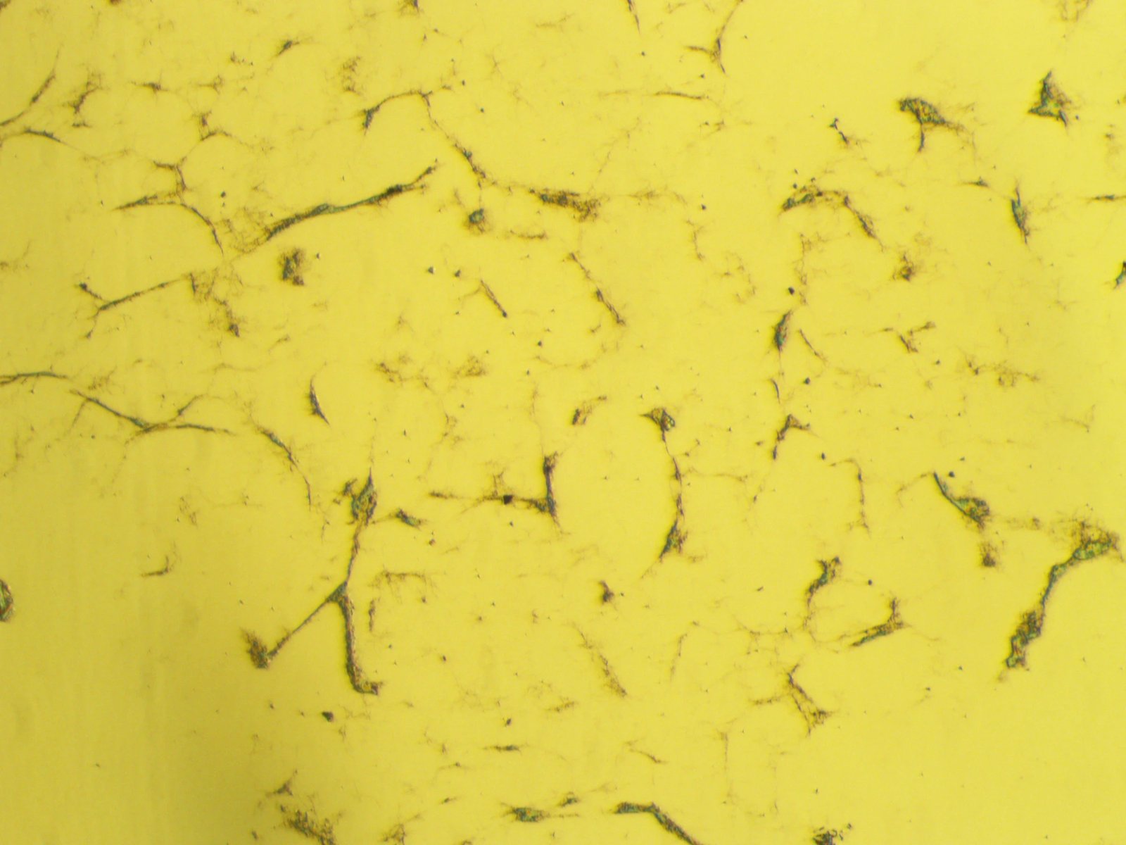
Optical, 10×: 100 µg/mL fibrinogen, 18 h on 50 µg/mL collagen. Only a sparse web of fine strands and small clusters, far less material than at 4 mg/mL. - Open question: are the fibres just the known hydrophobic-surface mechanism, or is the collagen film itself hydrophilic? → tested with contact angle on Day 8
Day 8 Wed 7/22/26
- Contact angle of collagen-only films (CAM 200 Optical Contact Angle Meter)
- Answers Day 7's open question
| Trial | Left angle | Right angle |
|---|---|---|
| 1 | 23.27° | 30.49° |
| 2 | 20.47° | 22.46° |
| 3 | 32.49° | 30.12° |
- Mean 26.55° (SD 5.06°, SE 2.07°)
- Well below the Berg limit of 65° → collagen film is hydrophilic, so fibres are not explained by hydrophobic adsorption alone
Day 9 Thu 7/23/26
- Deposited fibrinogen on 5 µg/mL collagen
- 1 h adsorption: 20 µg/mL and 100 µg/mL
- 18 h adsorption: 100 µg/mL
- Deposited 4 mg/mL fibrinogen on 50 µg/mL collagen, 18 h
- Stained 1 h samples with anti-Bβ122–134, then fluorescent secondary
Day 10 Fri 7/24/26
- 1 h adsorption, 100 µg/mL on 5 µg/mL collagen
- Imaged with optical microscopy and AFM
- Optical: 100 µg/mL, 18 h, on 5 µg/mL collagen
- AFM: 4 mg/mL, 18 h, on 50 µg/mL collagen
Day 11 Mon 7/27/26
- Canoe trip!
Day 12 Tue 7/28/26
- Reviewed AFM/optical results across collagen (5 vs 50 µg/mL) and fibrinogen (20 µg/mL–4 mg/mL) concentrations
- Planned controls (don't need all)
- Bare silicon + PBS only
- Bare silicon + fibrinogen
- Collagen-coated silicon + PBS only
- Collagen-coated silicon + fibrinogen + thrombin (positive control)
- Planned fluorescent staining: αC (fibrin structure, polymerization) and FpA/FpB (fibrinogen cleavage to fibrin)
- Antibody check: mouse anti-FpA (Novus NB100-73043, clone 49D2)
- Binds fibrinopeptide A (Aα1–16) on the fibrinogen α chain, plus free FpA
- FpA is cleaved off when fibrinogen converts to fibrin, so this stain marks fibrinogen and isn't specific to fibrin
Day 13 Wed 7/29/26
- Prepared confocal samples
- Collagen-only control, 50 µg/mL, collagen stain
- 4 mg/mL fibrinogen, 18 h, on 5 µg/mL collagen, mouse anti-FpA
- Each stain left to cover the wafer surface for 1 h
Day 14 Thu 7/30/26
- Confocal: collagen-only control, 50 µg/mL, two scales
- Confocal: 4 mg/mL fibrinogen, 18 h, on 5 µg/mL collagen, anti-FpA
- Imaged at 7 scales, including two Airyscan super-resolution images
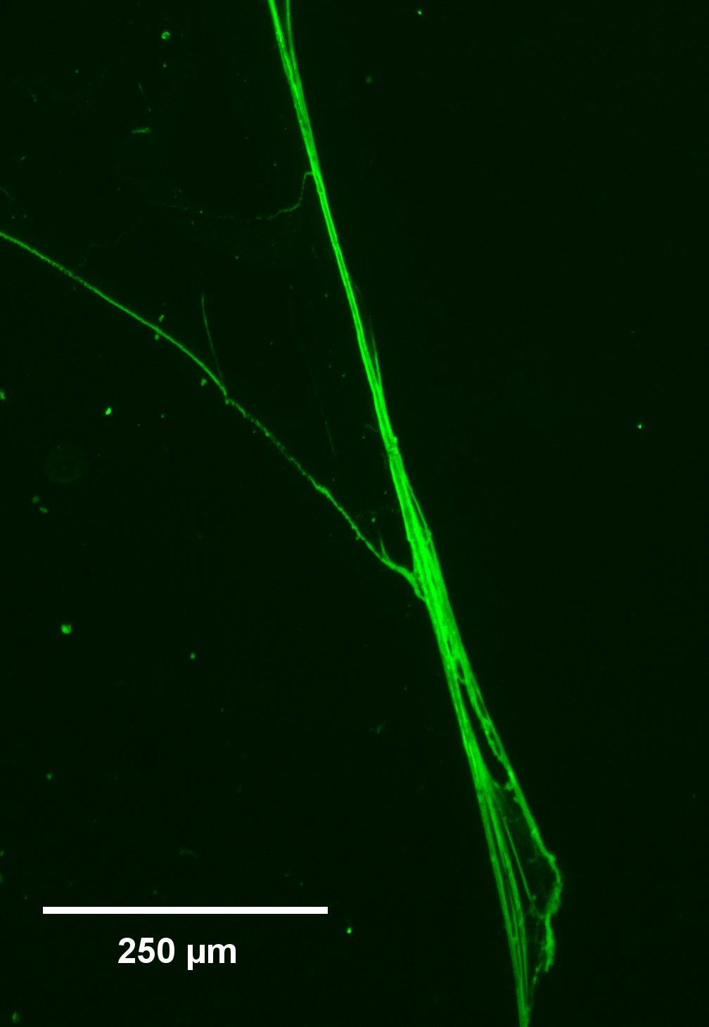
Widefield fluorescence (EVOS): 4 mg/mL fibrinogen on 5 µg/mL collagen, anti-FpA. Long bundled fibres light up along their whole length. Scale bar 250 µm. 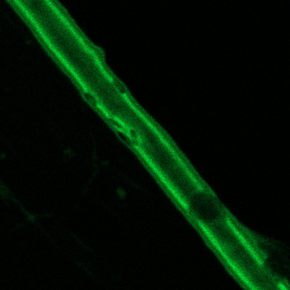
Confocal close-up of a single anti-FpA-stained fibre; the stain outlines the fibre along its length. (Day 15: this stain alone can't tell fibrin from fibrinogen.)
Day 15 Fri 7/31/26
- Analyzed confocal images; anti-FpA alone can't separate fibrin from fibrinogen → co-stain with anti-Bβ14–15 (knob B)
- Deposited fibrinogen on collagen for 18 h: 4 mg/mL for co-staining, plus samples for the αC time series
Day 16 Mon 8/3/26
- Co-stained 4 mg/mL samples, 1 h per stain
- Anti-Aα1–16 (anti-FpA) → fibrinogen (red)
- Anti-Bβ14–15 (anti-knob B) → fibrin
- Confocal: fibres are polymerized fibrin lying in cavities within a bed of fibrinogen
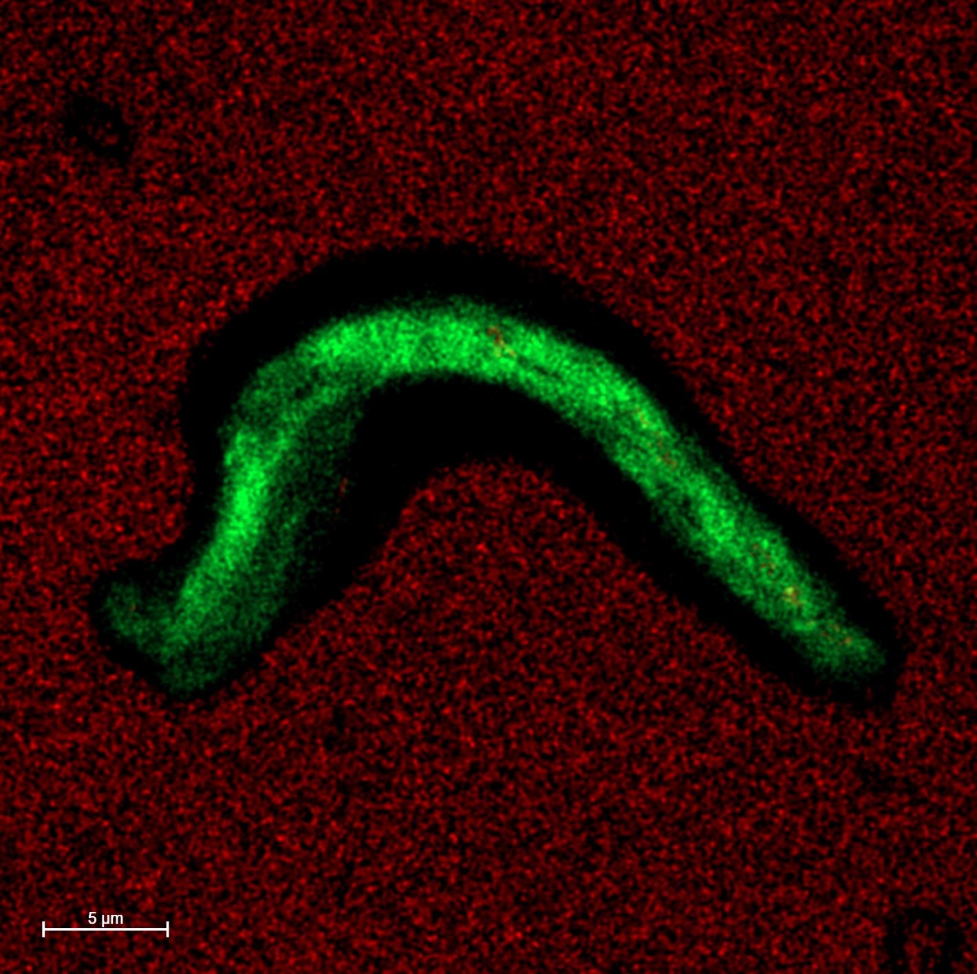
Confocal co-stain, 5 µm scale bar: a fibrin fibre (green, anti-knob B) lies in a dark cavity cut into the surrounding fibrinogen layer (red, anti-FpA). - αC time series: deposited matching 1 h samples; stained 1 h and 18 h samples with anti-Bβ122–134 (open αC)
- Fluorescence increased with adsorption time → layers of fibrinogen with open αC domains build up on collagen, similar to the hydrophobic-surface mechanism
- P12 setup: 50 µM P12 (fibronectin-derived antithrombotic, binds N-terminal subdomain of αC) mixed with 3.96 mg/mL fibrinogen, deposited on collagen for 18 h
Day 17 Tue 8/4/26
- Rinsed and imaged P12-treated samples vs untreated 4 mg/mL
- Reduced fibrillogenesis with P12 → assembly is mediated by the αC domain
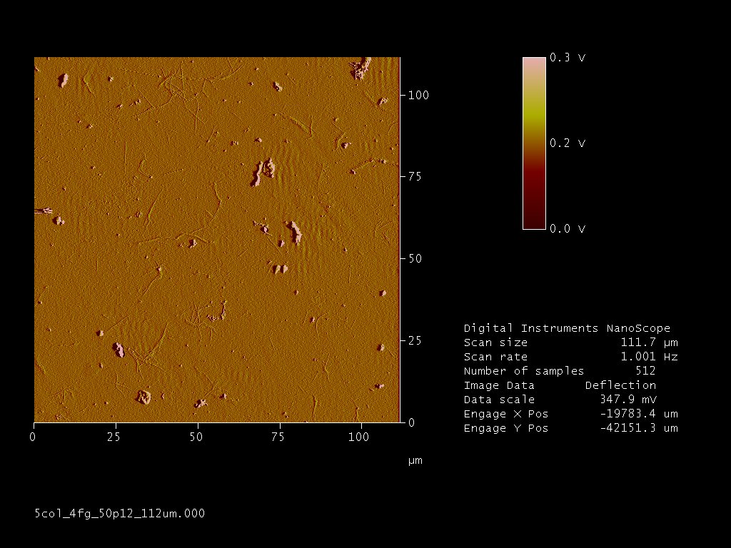
AFM, 111.7 µm scan: 4 mg/mL fibrinogen + 50 µM P12 on 5 µg/mL collagen, 18 h. Only short, thin fibrils and small clumps, none of the large fibres seen without P12. - Started presentation/abstract outline
Day 18 Wed 8/5/26
- Truncated αC assay
- 50 µg/mL fibrinogen des Aα414–610 and des Aα505–610, plus negative control, deposited 30 min on silicon
- 5 µg/mL collagen adsorbed on the fibrinogen films for 30 min
- Rinsed and stained with anti-collagen type I
Day 19 Thu 8/6/26
- Last images: confocal of truncated αC samples
- Anti-collagen type I intensity significantly lower on des Aα414–610 than on negative control and des Aα505–610
- → Aα414–505 (N-terminal subdomain of αC) is responsible for collagen adsorption, consistent with P12
- Finished presentation and extended abstract
Day 20 Fri 8/7/26
- Gave final presentation
Current Ongoing
- Accepted & presenting @ Materials Science Research Conference Dec '26 Boston conference for SB07, Emerging Materials for In Vitro Diagnostics
- Framing: malignant clot formation (stroke thrombi with lysis-resistant shells; collagen in clots impedes thrombolysis)
- Key points: thrombin-free fibrin on collagen (AFM); fibrin in a cavitated fibrinogen bed (confocal); αC untethering like hydrophobic surfaces; binding region Aα414–505; inhibited by P12
- Refining poster
- Writing paper for Journal of Thrombosis and Haemostasis